Hello! Today, we’ll be discussing cell injury and cell death, focusing specifically on the mechanisms, types, and causes of cell injury. The ultimate goal of every cell in the body is to maintain homeostasis. However, changes in the microenvironment can lead to structural and functional alterations, collectively known as cell adaptation. These changes can be:

- Physiological: Such as increased demand or hormonal influences.
- Pathological: Triggered by injurious agents like ischemia or toxins.
There are four types of cell adaptations: hypertrophy, hyperplasia, atrophy, and metaplasia,
When cells fail to adapt to their environment, they experience cell injury, which can be either reversible or irreversible:
- Reversible Injury: If the stimulus is removed, the cell can return to its normal state.
- Irreversible Injury: The cell cannot recover, even if the stimulus is removed, ultimately resulting in death by necrosis or apoptosis.
Causes of Cell Injury
The common causes of cell injury include:
- Hypoxia and ischemia
- Chemical agents
- Physical agents: Electric shock, radiation, and trauma
- Infectious agents: Viruses and bacteria
- Immunological reactions: Autoimmune diseases
- Genetic defects
- Nutritional imbalances
- Aging
Mechanisms of Cell Injury
Let’s explore the key mechanisms of cell injury:
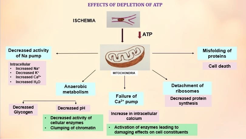
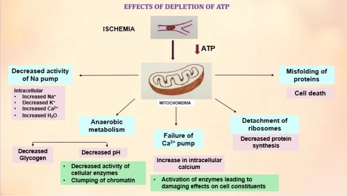
1. ATP Depletion
A decrease in ATP levels, often due to hypoxia or toxic injury, has several effects:
- Sodium-Potassium Pump Dysfunction: ATP is essential for maintaining electrolyte balance. Reduced ATP impairs pump activity, leading to sodium influx, water retention, and cellular swelling.
- Anaerobic Glycolysis Activation: Hypoxia increases glycolysis, leading to lactic acid buildup, reduced intracellular pH, and enzymatic dysfunction.
- Depletion of Glycogen Stores: Glycolysis consumes glucose from glycogen stores, depleting them over time.
- Calcium Pump Failure: Reduced ATP causes excessive calcium influx, which exacerbates injury.
2. Mitochondrial Damage
Mitochondria can be damaged by reactive oxygen species (ROS), hypoxia, and increased cytosolic calcium levels. Consequences include:
- Mitochondrial Permeability Transition Pores: These allow calcium ions to leak, disrupt oxidative phosphorylation, and reduce ATP production.
- Release of Apoptotic Proteins: Proteins like cytochrome C activate apoptotic pathways, leading to programmed cell death.
3. Calcium Influx and Loss of Calcium Homeostasis
Calcium levels are normally tightly regulated. Injury disrupts this balance, leading to:
- Activation of Harmful Enzymes:
- Phospholipases: Cause membrane damage.
- Endonucleases: Fragment DNA.
- Proteases: Break down proteins.
- ATPases: Hydrolyze ATP.
- Mitochondrial Damage: Formation of permeability transition pores and energy failure.
4. Oxidative Stress
Free radicals, highly reactive molecules with unpaired electrons, can damage cells via:
- Lipid Peroxidation: Damaging cell membranes.
- Protein Modification: Altering enzyme structure and function.
- DNA Damage: Causing mutations and strand breaks.
Cells counteract free radicals using antioxidants (e.g., vitamins A, C, and E) and enzymes (e.g., catalase, superoxide dismutase, and glutathione peroxidase).
5. Defects in Membrane Permeability
Damage to cellular membranes—plasma, mitochondrial, or lysosomal—leads to:
- Plasma Membrane Damage: Disrupts ionic balance and causes cellular swelling.
- Lysosomal Rupture: Releases enzymes that digest cellular components.
- Mitochondrial Membrane Damage: Formation of transition pores and loss of energy production.
Features of Reversible Cell Injury
Reversible injury is characterized by cellular swelling and fatty change:
- Cellular Swelling:
- Increased cell size.
- Fluid-filled vesicles in the cytoplasm.
- Mitochondrial swelling, ribosomal detachment, and membrane abnormalities.
- Fatty Change:
- Abnormal accumulation of triglycerides, commonly in the liver.
- Seen in metabolic syndrome, toxin exposure (e.g., alcohol), and ischemia.
Visual Features
Microscopic features include:
- Swollen Cells: Enlarged cells with distended organelles.
- Myelin Figures: Lipid masses derived from damaged membranes.
- Chromatin Clumping: Due to increased acidity.
- Fatty Change in Tissues: Common in the liver, myocardium, and renal tubular cells.
That concludes this discussion on cell injury! In the next, we’ll delve into cell death mechanisms, including necrosis and apoptosis.

